ABSTRACT
Introduction. Increased overjet is a significant risk factor for dental trauma. Children with increased overjet are prone to incisor trauma in both deciduous and permanent dentitions. Case reports. Case 1: Girl (age 2y4m), brought in emergency after trauma, with complete intrusion of 51, subluxation of 61, increased overjet favoured by finger sucking. Extraction of 51 was performed after 3 months with no re-eruption. Myofunctional treatment of overjet was started at age 3y4m. After 6 months of myofunctional therapy overjet is significantly reduced, oral breathing and finger sucking ceased. Case 2: Boy (age 9y 4m) sought treatment for enamel-dentin crown fractures of 11 and 21, 3 weeks after trauma. Clinical examination shows pulp exposure in 11 with diminished response to vitality test, class II with increased overjet and oral breathing. Anamnesis revealed repeated traumatic episodes and history of finger sucking. Treatment started with pulpotomy in 11 and crown restorations in both incisors. During the following 6 months endodontic treatment had to be performed in both fractured teeth due to complications. Four subsequent traumatic episodes occurred during this time. Parents agreed to start orthodontic treatment 7 months after first visit. After 17 months of treatment (mobile followed by myofunctional), evolution is good, with aligned teeth, significantly reduced overjet, nasal breathing and no further traumatic injuries. Conclusions. Management of increased overjet in deciduous dentition is feasible and may help reduce risk of trauma in both dentitions at low costs. Postponing may result in permanent damage of permanent incisors and more complex treatment.
Key words: overjet, trauma, interception, myofunctional therapy
Traumatic dental injuries (TDI’s) have an important biological, psychological and physical impact in patient’s life, especially if they occur at a young age. They can affect both the deciduous and the permanent dentition, especially in the anterior part of the maxillary arch, the most common traumatized teeth being the maxillary central incisor (Luca 2013) [1], (Rajab 2013) [2], Rouhani (2014) [3], but their presence is more commonly identified in children under 12 years of old (Andersson 2013) [4]. Luca (2013) [1], Arraj (2019) [5], and Born (2019) [6] state that the risk of frontal dental trauma increases when the spaces between the two maxillas (the overjet) grows. TDI’s frequently occur in young patients with dento-maxillary anomalies, especially children with class II division 1 syndrome – the most common anomaly of the development of the dento-maxillary apparatus in our country (Boboc, 1971) [7] – which affects about 15 – 20% of the Caucasian population (Mitchell, 2013) [8]. Class II/1 malocclusion is characterized by insufficient transverse development of the dental arches, especially the maxillary arch, with important facial changes, as well as functional disorders (Boboc, 1971) [7]. Other features may be an open bite, dental protrusion or lip incompetence (Luca 2013) [1], (Cortes 2001) [9], but the most obvious clinical sign in class II/1 syndrome is the increased overjet, an important predisposing factor for TDI’s.
Some authors (Arraj,2019) [5] found a direct association between dental trauma and increased overjet; in deciduous dentition, an overjet greater than 3 mm comes with a risk of three times more TDI’s than a normal overjet, and in permanent dentition, an overjet greater than 5 mm may double the risk of dental traumas as compared to a well developed frontal occlusion (with 0 – 3 mm overjet). Born (2019) [6] points out that overjet and lip incompetence are important risk factors for dental trauma in deciduous dentition, and if overjet is greater than 3 mm, the risk of TDI is almost four times bigger. Antunes (2015) [10] found that children with different types of malocclusion have a 64% higher chance for TDI’s, patients with increased overjet being three times more exposed to dental trauma than kids with any other malocclusion. Tümen (2011) [11] states that risk of dental trauma in primary dentition is higher if the patient has an overjet greater than 5 mm, has an open bite or is a boy. Similar results on the relationship between increased overjet and TDI’s are reported by Dosdogru (2019) [12], Rajab (2013) [2], Rouhani (2014) [3] and Shakuntala (2019) [13].
For children with (or without) increased overjet who practice different types of sports considered at risk (wrestling, rugby etc), the use of mouthguards may help avoid dental trauma, as suggested by Knapic (2007) [14] and Mark (2017) [15]. Given the high prevalence of TDI’s, especially in children under 12 years of age (Andersson) [4], proper educational programs appear necessary in order to enhance general knowledge on preventing and managing these situations(Born) [6]. The Romanian National Association of Paediatric Dentistry (ANSPR) in cooperation with and Colgate Romania initiated a project in this respect (https://pedodontieromania.wixsite.com/anspr/reviste). With the contibution of the Romanian Red Cross, this initiative is expected to spread countrywide in the near future.
Although there is a direct relationship between dental trauma and increased overjet, there is still debate regarding early versus late orthodontic treatment in class II division 1 cases. Batista (2013) [16] conducted a study comparing early two-phase treatment (for patients between 7 and 12 years old), to late one-phase treatment (for patients aged 12 to 16 years) in patients with prominent upper teeth and increased overjet. The early treatment group (with functional appliances) had a lower incidence of incisor trauma compared to the late treatment group. Only 19% (31 out of 161) patients which had had early treatment reported recurent dental trauma, as compared to 30% (51 out of 171) patients with late treatment. Patients that had used headgear as an early treatment reported less new DTI’s (24 out of 117 patients), while in patients with late orthodontical treatment the proportion was double. As such, interceptive orthodontic treatment can be used to reduce the increased overjet and can help minimize the risc of TDI’s (Batista, 2013) [16]. Provision of early/interceptive orthodontics in children with increased overjet is more effective in reducing the incidence of traumatic dental injuries than orthodontic treatment during adolescence (Arraj) [5].
However, Abbot (2018) [17] considers that TDI’s need not be orthodontically intercepted during primary dentition and, if the overjet is greater than 3 mm, parents should made aware about the risk of the TDI’s and potential consequences, and children should be kept under observation. Only after the permanent central incisors erupt and if the overjet is greater than 5 mm, interceptive orthodontics can be sought in order to reduce the overjet and the risk for TDI’s. According to the National Health Service of England’s selection criteria, patients under the age of 18 years with an overjet greater than 3 mm qualify for orthodontic treatment supported by the NHS only if the increased overjet is associated with other dento-maxillary abnormalities, leading to a moderately unattractive dental appearance [18].
Given the relationship between increased overjet and the risk of TDI’s, it’s important to know how increased space between the two jaws occurs and what are the causes that may induce or enhance this increment. Boboc (1971) identified environmental factors (like mother’s diet during pregnancy and child feeding habits) with major influence on the occurence of class II division 1 malocclusion, yet heredity is not to be overlooked. Functional factors like thumb sucking and oral breathing may play a crucial role in enhancing if not determining the anomaly. Thumb sucking can produce proclination of the upper incisors, retroclination of the lower incisors, an open anterior bite or a lowering of the overbite and may lead to a narrowing in the upper arch by the negative pressure generated by this habit (Mitchell, 2013) [8]. All these consequences are determined by the duration and the intensity of the habit. Oral breathing acts by causing a decrease of air pressure in the nasal cavity and maxillary sinuses, letting the athmospheric pressure prevail; by permanently maintaining the mouth open, muscles will act on the lateral sectors of the maxilla and will limit its transveral development; the upper lip becomes short and hypotonic and will allow the upper teeth to tilt foward [19]. These effects will be superimposed upon the child’s existing developmental pattern and thus can lead to an increased overjet and a retruded mandible (Mitchell, 2013) [8].
Given the facial changes in class II division 1 anomalies and the high risk of traumatic dental injuries that may occur in both deciduous and permanent dentition [19, 8], some authors recommend orthodontic interception as soon as the first signs of abnormal development appear [10]. Functional appliances have been used in orthodontics since the 20th century to correct Class II malocclusion. DiBiase (2015) states that “clinical effectiveness of these appliances is acknowledged and they can be very useful in the correction of sagittal arch discrepancies”[20]. Positive effects on skeletal, dental and profile aspects by using functional appliances in the interceptive orthodontic treatment of Class II/1 malocclusions were also noted by de Bittencourt in 2015 [21]. Franchi (2013) [22] found that the use of functional appliances for this type of malocclusion was associated with improvements in overjet management. Myofunctional orthodontic treatment uses mobile devices to correct the altered functions that caused or enhanced the malocclusion while the child is still growing. Myofunctional orthodontics tonify orbicular muscles, correct the position of the tongue, ensure physiological nasal breathing, balance muscles and favour teeth alignment within the muscle matrix [23]. Performing myofunctional treatment requires a good understanding of the role of altered functions within the dento-maxillary abnormalities. During physiological growth, the facial massive develops forward and downwards, providing the space needed to align the anterior and posterior teeth as they erupt [24]. If facial muscles work within normal limits, the tongue is in the correct position (behind the incisive papilla), breathing is done through the nose and the patient swallows with the lips in contact, harmonious growth occurs [19]. If the patient is an chronic oral breather, the mouth is open most of the time and the tongue position is low. Under the action of the imbalanced intraoral and perioral muscles development becomes more vertical than horizontal and changes occur in both arches (the maxilla narrows and crowding occurs) and in the development of the cephalic extremity (mandible will set to a posterior, retrognat position, with a convex profile) [19].
There are a number of factors that need to be considered when seeking myofunctional treatment, some of which are the patient’s constitutional pattern, the motivation of patients, and the desire for treatment [22].
Two clinical cases of class II/1 malocclusions with increased overjet and vicious habits (oral breathing and thumb sucking) are presented below. In both cases (one with deciduous and the other with mixed dentition) the reason for seeking dental treatment was the same – traumatic dental injuries.
CASE I
Girl, 2 years and 4 months old, was brought in emergency with dental trauma. Clinical examination revealed complete intrusion of 51, subluxation of 61 and increased overjet (Figure 1).

a 
b 
c


The patient was a chronic oral breather and anamnesis confirmed that she consistently performed finger sucking.
For the following 3 months no spontaneous re-eruption was seen. Therefore, non-traumatic extraction of 51 was performed under nitrous oxide sedation and local infiltration.
Vicious habit and oral breathing, as well as the increased overjet, were addressed by myofunctional orthodontic treatment with MyobraceR trainer as soon as parents felt that reasonable cooperation was to be expected from the child – at age 3y 4m (Figure 2).

a 
b 
c 
d


After 6 months of myofunctional therapy (MyobraceR) overjet is significantly reduced, oral breathing and finger sucking ceased, and the patient is happy with her new appearance (Figure 3).

a 
b 
c


CASE II
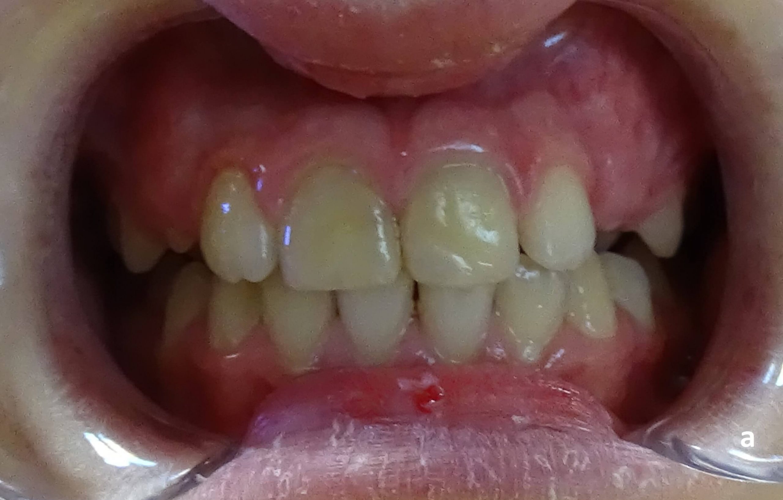
Boy, 9 years and 4 months old, sought treatment for extensive enamel-dentin crown fractures of 11 and 21, 3 weeks after dental trauma occured. Clinical examination revealed pulp exposure, modified colour and diminished response to vitality test of the upper right central incisor (Figure 4 a, b). 21 was still vital, with no pulp exposure. Class II malocclusion with increased overjet and crowding in both upper and lower jaw were identified. The patient was a chronic oral breather.

a b 
c


Anamnesis revealed that the patient had a history finger sucking, with repeated mild traumatic episodes. The general practitioner had recommended orthodontic examination in the past.
Treatment began by a pulpotomy in 11 (Fig. 5a). During the following 6 months, 4 subsequent trauma episodes occurred. Endodontics with partial pulpectomy (MTA) had to be carried out in both fractured teeth due to complications (Figure 5, a-c).

a 
b 
c


Parents agreed to start the orthodontic treatment 7 months after first visit (Figure 6).

a 
b


After 17 months of orthodontic treatment (mobile followed by myofunctional – with a MyobraceR trainer device) evolution is good, with aligned teeth, significantly reduced overjet, nasal breathing and no further traumatic injuries (Figure 7).

a b

Conclusions
- Early management of increased overjet in deciduous dentition is feasible and may help reduce the risk of dental trauma, both in temporary and permanent dentition, at minimal costs but maximum efficiency for a harmonious development of the patient.
- Postponing the orthodontic treatment may result in traumatic injuries in permanent upper incisors and need for more complex treatment.
- Interceptive orthodontics by myofunctional therapy can be an effective way of limiting the aggravating evolution of the malocclusion and preventing traumatic dental injuries in young patients.
REFERENCES
- Luca R. – Pedodonție Volum 3, Editura Cermaprint, București, 2013: 5 – 64
- Rajab LD. et at. – Traumatic dental injuries among 12-year-old schoolchildren in Jordan: prevalence, risk factors and treatment need, Oral Health Prev Dent. 2013;11(2):105-12.
- Rouhani A. et al. – Anterior Traumatic Dental Injuries in East Iranian School Children: Prevalence and Risk Factors, Iran Endod J. 2015 Winter; 10(1): 35–38.
- Andersson L. Epidemiology of traumatic dental injuries. J Endod. 2013; 39: S2– 5
- Arraj G.P. et al. – The association of overjet size and traumatic dental injuries—A systematic review and meta‐analysis, Dental Traumatology 2019; 217 – 232
- Born C.D. et al. – Traumatic dental injuries in preschool‐age children: Prevalence and risk factors, Clin Exp Dent Res 2019 Apr; 5(2): 151–159.
- Boboc G. – Anomaliile Dentomaxilare, Editura Medicală, București, 1971: 65 – 91
- Mitchell L. – An introduction to orthodontics. Fourth Edition. Oxford University Press, 2013: 114-125
- Cortes, M.I., Marcenes, W. and Sheiham, A. (2001): Prevalence and correlates of traumatic injuries to the permanent teeth of schoolchildren aged 9–14 years in Belo Horizonte, Brazil., Dental Traumatology 17, 22–26.
- Antunes L.A.A. et al. – Increased overjet is a risk factor for dental trauma in preschool children. Indian Journal of Dental Research (2015). 26. 356. 10.4103/0970-9290.167630.
- Tümen et al. – Incisor trauma in a Turkish preschool population: Prevalence and socio-economic risk factors, Community Dental Health (2011) 28, 308–312
- Dosdogru Y. et at. – Maxillary incisor trauma in patients with class II division 1 dental malocclusion: associated factors. Journal of Istanbul University Faculty of Dentistry 2017;51(1):34.
- Shakuntala.B.S, Amruta Kalpavriksha – Prevalence Of Traumatic Dental Injuries Among Residential School Children And Day Scholars Of 9-14 Years In South Bangalore, International Journal of Scientific and Research Publications, Volume 9, Issue 8, August 2019
- Knapik et al. – Mouthguards in sport activities: history, physical properties and injury prevention effectiveness. Sports Med. 2007;37:117–44.
- Mark A.M. – Keeping your teeth and mouth safe. J Am Dent Assoc. 2017;148: 476
- Batista K.B. et al. – Orthodontic treatment for prominent upper front teeth (Class II malocclusion) in children and adolescents, Cochrane Database Syst Rev. 2018 Mar 13;3.
- Abbott P.V. Traumatic dental injuries are now the 5th most prevalent disease/injury in the world – But they are being neglected!!, Dental Traumatology 2018;34:383.
- NHS England.Guide for Commissioning Orthodontics 2015.
- Boboc G. – Aparatul Dento-Maxilar, Formare și dezvoltare. Ediția a II-a, Editura Medicală, București, 2016: 317 – 30, 433 – 45
- DiBiase A.T. et al. – The use of functional appliances in contemporary orthodontic practice, British Dental Journal 2015 Feb, 16;218(3)
- de Bittencourt Neto AC et al. – Therapeutic approach to Class II, Division 1 malocclusion with maxillary functional orthopedics, Dental Press J Orthod. 2015 Jul-Aug;20(4):99-125.
- Franchi L et al. – Long-term skeletal and dental effects and treatment timing for functional appliances in Class II malocclusion, The Angle Orthodontist 2013 Mar;83(2):334-40.
- Homem MA et al. Effectiveness of orofacial myofunctional therapy in orthodontic patients: A systematic review. Dental Press J Orthod. 2014 July- Aug;19(4):94-9.
Premkumar S., Textbook of Craniofacial Growth, Jaypee Brothers Medical Publishers, New Delhi, India, 2011: 86-105


